How Accessible Publishing has Revolutionized Academic Publishing

The world of scientific publishing has undergone a metamorphosis in recent times. The key ingredients here are “authentication” and “piracy” in scholarly communications. Many people have come up with “inclusiveness of scholarly communications,” for disabled people. There seems to be a lacuna in the world of scientific publications, given the “professional” and “educational” spheres of education. In a highly connected world, knowledge must be disseminated through “journals,” “books,” and “databases.”

At one end, most academicians are of the view that all knowledge must grow with highly authoritative communications being accessible to readers. At the other end, publishers argue about “copyright infringement” with new challenges towards tackling piracy and digital infringement. Accessibility is still an issue for scholarly communications.

According to the National Institute of Health, there are more than 285 million people with some form of physical, cognitive, and educational challenge in the USA. Nevertheless, the enrollment of disabled masses is just 10–20% across colleges in the USA. People with disabilities are a sizeable population if we take into consideration that they can be perennial customers of scholarly communications.

“Accessibility” is still an issue here for most publishers. Publishers are concerned about “return on investment” while catering to such people. Accessible publishing is not really avenue for making great revenues. However, people with disabilities are now being considered for inclusiveness and diversity. They are provided with “navigable, feature-rich content” through various innovative publishers: DAISY and ReadSpeaker.

Accessible publishing is far superior given the fact that it can improve the “quality” and “interoperability” of metadata. With proliferation of machine learning and search engine optimization (SEO), general discoverability of such innovative publications is improved significantly. Publishers and technological evangelists provide deciphering workflow with maximum accessibility to all stages of publications, right from manuscript selection, improvement, and publications. Right from increasing submissions to improving usage, measurable benefits can be availed through “accessible publishing.”

Accessible publishing is much easier today thanks to developments in technology although it does require some efforts out here. Technologies such as HTML, EPUB, etc., would be leveraged to provide the best practices in accessible publishing. Industry-standard workflow formats of publishing are now accessible to all readers. The mission of academic publishing would be to disseminate knowledge through “accessible publishing.” They can envision the horizon of many accessible publishers.


In clinical drug trials of heart disease, women and older patients are under-represented

Doctors have to refer to randomized clinical trials to determine the best ways of treating patients.  Furthermore, doctors refer to this information to also determine the most suitable drug that can be prescribed to these patients. Heart illness is the most common disease that afflicts the common man. In recent years, it has been proven that the number of women having heart ailments would be greater than the number of men having the same ailment. Compared to younger people, older people have a greater tendency of developing heart condition. Does the data presented in clinical trials actually exhibit reality?

In most cases, the data does not actually represent the true picture. A new study was recently published in the journal Circulation: Cardiovascular Quality and Outcomes. Professor Quoc Dinh Nguyen works at Université de Montreal’s Faculty of Medicine. He supervised a team of researchers who tested new heart drugs on mostly men (71 per cent); however, the majority of people afflicted with heart disease were mostly women. Moreover, the average age of male patients with heart disease was 63; however, the average age of patients who suffered the two most common heart diseases was in the range of 68–69 years.

In the past 20 years, the gender and age gap between subjects participating in drug trials has hardly diminished; however, the population seems to be aging rapidly. Professor Quoc Dinh Nguyen is a geriatrician who works at the Centre Hospitalier de l’Université de Montréal (University of Montreal Hospital Center).

In most drug clinical trials, both women and older patients are under-represented; therefore, both these groups of patients would receive comparatively lesser care. Unlike a young patient, an older patient does not really respond much to several treatments and medications. Many-a-times, it is very difficult for patients to receive an exact dosage or intervention; moreover, each medication has a set of severe side-effects.  However, we do not know the specific course of medication until and unless a large number of older patients are included in clinical trials. Most studies also do not include women in their clinical trials.

In the present scenario, the findings are obtained from a clinical trials conducted predominantly on male and younger subjects. These results do not really patient outcome of women and older subjects generally. Nguyen examined the issue closely while working as a geriatric resident. They discussed with colleagues to realize that heart disease is a grey area to receive effective treatment. Resident physicians of other departments (anaesthesiology, psychiatry, emergency medicine, and cardiology) also collaborated in their efforts of improving the results.

A brief history

Nearly 20 years ago, researchers were concerned about how under-represented were several sections of the society, especially women. The results of clinical trials were quite often problematic in nature. With a team of researchers headed by Nguyen, we set off to find out if these practices had improved significantly. The 25 most frequently cited clinical trials were examined closely every year. The examination period was of twenty years, ranging from 1996 to 2015. The data was published in the U.S National Health and Nutrition Examination Survey 2015-2016; this data compared how prevalent was cardiovascular disease in America . The  data was classified in terms of following parameters: age and gender.

The research team examined data of following medical conditions: coronary artery disease, hypertension, heart failure, atrial fibrillation. This team also examined several risk factors contributing to cardiovascular diseases. This research team closely examined the correlation between diabetes and heart disease. Previous studies have reported that diabetic patients were more likely to suffer from coronary heart disease.

Bad results

Currently, a greater number of women and older patients are included in clinical trials; therefore, there has been a slight improvement in the representational bias of clinical trials.   Eric Peters works as an anesthesiologist at the CHU Saint-Justine children’s hospital; he is the second author of this study. Depending on our calculations, it would take another 90 years to understand whether clinical trial studies could present data correctly without bias. We need to correctly understand the factors contributing to coronary heart disease.  The factors leading to aging population must also be considered in this situation.

After analyzing 500 clinical trials, we arrived at the following conclusion: only 29 percent of participants were women in this clinical trial. Moreover, the average age of participants was just 63 years. According to Nquyen, the reality is quite different in the hospital emergency rooms and departments; these departments are of internal medicine, cardiology, and geriatric medicine. Women and older patients were hardly represented in clinical trials that were focused on determining the factors associated with CAD and heart failure. Women represent more than 54.6 percent of CAD patients. In clinical trials, more than 27.4 percent of participants for CAD were women.

Is heart disease really a man thing?

It is a general perception that men are afflicted with heart disease; however, most medical research studies have reported about results that are completely obsolete. Heart disease is the leading cause of women’s death in Canada. Fewer men die of heart disease in Canada. In general, heart disease would affect women at a later stage in life. Nearly after 10 years, women would die of coronary artery disease and heart failure. There has been steady decline in the death caused by heart disease in women; moreover, death caused due to heart disease would be greater in men. One of the most common hypothesis is the fact that men receive timely medical treatment unlike women.

Why are women excluded from clinical trial?

In general, most women were excluded from clinical trials because it was advisable to give them medication during pregnancy; however, this principle should not have been applicable to drugs used for treating heart condition. This is because most patients with heart conditions are usually more than 60 years of age. To select a woman to participate in clinical trial, we also need to consider the age of the woman. To ensure the adequate participation of women in clinical trials, older patients should be recruited. This is because women are afflicted with cardiovascular disease at a comparatively later stage of treatment, unlike men.

In general, it is difficult to conduct clinical trial of older patients. This is because most older subjects would find it difficult to move around; moreover, it is usually tougher for them to undergo a battery of clinical tests. Older the patients, higher would be their difficulty in moving around.  In general, older patients do require several medications as they are afflicted with several ailments.


The impact of White Lies in Technological Era

The Journal of Language and Social Psychology states that most Americans would speak lies once or twice each day. However, we have a tendency to take truth as face value, intending to accept dishonest replies as honest answers to questions. Our lie-detecting skills are completely vandalized, raising issues of transparency. These capacities would be completely detrimental to one’s discerning capacity.

Coming back to how it affects the publishing arena, let’s just say that most budding researchers would look forward to the opportunity of reading an entire manuscript as a peer reviewer; however, quite a few peer reviewers are voluntary workers who just skim through the manuscript. In the sense, they were “close enough” to reading an entire manuscript.  How do we judge the integrity and authenticity of latest work? Let’s just say that it is absolutely difficult to decipher the quality of work. In the course of their professional careers, most researchers would be involved in writing, editing, and research. This is particularly true for people who need to their careers in scientific publishing. The concept of “white lies” has eroded the integrity of publishing industry.

Technological “white lies”

Consider the recent scandal of data mining from Facebook. The user agreement of this website claims to keep all data secure; however, internet is an online medium of shady grey areas. With no internet censorship, there are many shady dens operating on making fake profiles that not just abusive but also derogatory. Data is rigged and sold to most of these companies as nothing really is hack-proof in the “internet of things.” What really gets people hooked or compelled to Facebook? Does the website really guarantee privacy of information? Nope. The truth absolutely seems to be presented as a “white lie.”

Healthcare “white lies”

With the internet of things, we have now revolutionized the concept of healthcare advertising. With herbal and non-conventional medicines, there are umpteen videos claiming to restore chronic health ailments through various herbal formulations. Do these agencies really get approved by FDA? Nope, but the fine print is often hidden to mask the ignorance of general consumer. How much of the work done impact the industry of healthcare publishing on a global scale?

Marketing copywriters in the pharmaceutical industries don’t really have any direct impact on the integrity of pharmaceutical data; however, there are quite a few medical researchers, academics, and scholars who will have a dent with a slightly inaccurate data being presented. Their scholarly, influential outputs make a “yes” or “no” decision in the drug-regulating authorities.

Media “white lies”

Many things matter in the age of digital media. Fake news sells like hot cakes these days as practically every video can be morphed and altered to sabotage the reputation of your contender. These videos are then circulated through databases to cause riot like situation. The legal authorities use this situation for their vested interests and gain mileage. Does anyone really go out of limits to censor these fake news videos? Nope. Internet should never be considered as “free speech” but the lobby of Silicon Valley is too weighty to break off.

Plagiarism is being unraveled in the publishing industry like never before. The recent scandal of plagiarism and hacking has shaken the integrity of large publishing houses: Elsevier, Springer Nature, and Wiley. “Hacking” and “identity theft” are the newest buzzwords in the technological world. Has anyone really checked the integrity of the so-called digital marketing agencies, claiming to have been managing the “online reputation” for a whole set of fake profiles, netizens, sleaze, and slander. Nope. Photoshop is an imaging software used for producing doctored images. Do modern businesses follow any rules of ethics? Nope. Why? That’s because the lobby of sinister pigs flourishes under the garb of “internet is free speech.” The term “cyberterrorism,” “cyberabuse,” are just used to mask these disgusting, menacing situations.

Conclusion: White lies have become the conventional method of sensationalizing things and vandalizing integrity. With technological evolution, better tools have been developed to catch crooks but there are also many technologies that are being used by powerful crooks to sabotage the system.


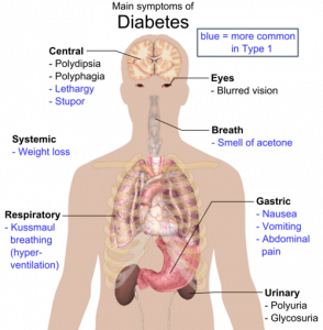
Verapamil: an effective therapy for type 1 diabetes

To promote the functioning of beta cells and insulin in patients with type 1 diabetes, researchers have developed a novel strategy that minimizes the requirements of insulin and the incidence of hypoglycemia. These researchers have worked at the Comprehensive Diabetes Center at the University of Alabama in Birmingham, USA.

The journal Nature Medicine published these findings recently. Verapamil is the most commonly used drug for controlling blood pressure; it was approved for oral administration in the year 1981. Following the administration of verapamil, type 1 diabetes patients were able to produce greater amounts of insulin. Thus, their daily requirement of insulin was reduced substantially and their blood sugar levels were also in control. The drug verapamil is not just safe and effective for type 1 diabetes but also a promising therapy that provides new hope to people living with this life-threatening condition. These were positive results confirmed in a human clinical trial that was randomized and double-blinded in nature; the clinical trial was controlled by placebo.

Verapamil has shown promising results in improving the function of beta cells in pancreas; the functioning of beta cells is related to the control of insulin production. Optimum levels of insulin ensure a good quality of life in patients. Under such a scenario, type 1 diabetes patients have new hope. It is otherwise difficult to control such a life-threatening condition that offers no hope. Although this drug does not sound like a complete cure for type 1 diabetes, it is however a promising therapy for altering a life-threatening condition: type 1 diabetes. Such patients need to boost the production of insulin in their body in order to have better disease control.

A clinical trial was conducted on animal models in the year 2014. In this clinical trial, it was reported that the condition of type 1 diabetes could be completely reversed by administering verapamil. Then, they conducted a human clinical trial to determine the effects of this drug. For more than three decades, the drug verapamil has been approved by the FDA for the treatment of high blood pressure. Current research findings are path-breaking in the sense that the drug is quite safe and effective in the treatment of type 1 diabetes patients. In patients with type 1 diabetes, the body’s immune system attacks beta cells of the pancreas. These beta cells are responsible for the production of insulin. Insulin is the hormone that controls blood sugar levels in the patient.

The production of insulin decreases substantially when the beta cells of pancreas are destroyed in the human body. Consequently, blood sugar levels would rise in the human body and the patient would become extremely dependent on external sources for insulin. The function of beta cells can be preserved effectively when a patient is administered verapamil. This drug induces the body to produce more insulin. In various clinical trials, it has been proved that the participants’ dependency on external insulin decreases substantially. Several individuals with type 1 diabetes can effectively regulate their blood sugar levels with this strategy.

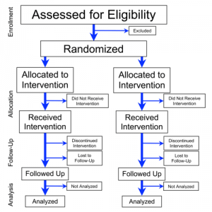
In the human clinical trial, the drug verapamil was administered to 24 patients. These patients were in the age group of 18 to 45 years. Over the course of one year, verapamil was administered to 11 patients while a placebo drug was administered to 13 patients. Only patients with type 1 diabetes were included in this clinical trial. They received insulin therapy to manage their condition throughout the duration of this clinical trial. The total daily dose of insulin was monitored in both the groups, that is, the group that received verapamil and the group that received placebo. Moreover, we also monitored the amount of insulin produced in these groups. Factors such as the percentage of change in insulin and HbA1C levels were also monitored. There were patients who experienced hypoglycemic events; all such events of each patient were recorded in our human clinical trial. A continuous glucose monitoring system was used to determine the healthy blood glucose levels of each patient.

Patients with type 1 diabetes do have therapeutic options for hope. In fact, such patients should be able to deal with the illness in a promising way following the successful administration of verapamil. Insulin dependency was substantially reduced in patients with type 1 diabetes following the administration of verapamil. The quality of life was significantly improved in these patients. The risk of comorbidities would be improved when the overall blood sugar levels was controlled in patients. Thus, a patient with type 1 diabetes would not develop several other comorbidities, such as kidney disease, blindness, and heart attack.


Cancer stem cells can now be destroyed by targeting metabolism

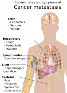
Cancer is a fatal illness with poor prognosis and survival rate, especially when it has progressed to a metastatic state. Scientists have not yet been able to decipher why patients become resistant to chemotherapeutic drugs and therapies. To address this objective, researchers worked diligently at  the Rogel Cancer Center—it is affiliated to the University of Michigan. They made an important breakthrough in the year 2003. The lead supervisor was Dr S. Wicha, MD for the team of researchers. They found that there are cancer stem cells that act like a fuel within a tumor. Although this group of cells is immensely small, they are the ones that trigger the growth and metastasis of cancer. The simple strategy was then to simply kill the group of cancer stem cells, and the long lost battle against cancer could be defeated easily. But, is this so easy to sound hopeful for cancer patients? Not really, cancer is such a condition that can relapse and attack patients even after they have been cured temporarily.


Currently, there has been an important discovery: cancer stem cells do not really exist in ONLY a single state but they are exhibited in different states; they are immensely plastic in nature. This implies that different forms can be easily adopted by cancer stem cells. They could be in a dormant state for some point of time and then easily bounce back into uncontrolled growth, leading to formation of tumor. Multiplication and spreading, the two characteristic features of cancer stem cells, have been attributed to its most important property: plasticity.

Presently, patients are treated with targeted therapies for combating cancer. Although these therapies are effective, they have been successful in destroying tumor cells only for a certain period of time. There are many cases in which patients develop resistance to these targeted therapies. What is the cause of drug resistance in cancer patients? Most scientists believe that drug resistance is triggered once again by cancer stem cells. Because cancer stem cells have high plasticity, they change their form completed when subjected to targeted therapies. The resultant effect is that cancer stem cells are completely unrecognizable to these therapies following change of form. The patient thus develops resistance to therapies and the patients’ condition deteriorates consistently.

The conclusion: multiple stem cell therapies must be developed to effectively combat every form of cancer stem cell. This is a humungous task to achieve according to scientists at the Rogel Cancer Center. Cell metabolism is the key feature that controls the plasticity of cancer stem cells. How do we eliminate the plasticity of cancer stem cells? Well, all we need to do is to target the metabolism of cancer stem cells. In other words, cancer stem cells can be effectively attacked by destroying cell metabolism.

Mitochondria are cell organelles that supply energy to cells, irrespective of its kind. This includes cancer stem cells. Mitochondria are organelles that perform cellular respiration, depending completely on the supply of oxygen. Cells derive energy from mitochondria, which converts sugar or glucose molecules into energy with the help of cellular oxygen. Cancer stem cells are very unique due to its plasticity. When they are in the dormant state, they derive energy from glucose molecule. When they grow in a proliferative state, cancer stem cells depend completely on oxygen.

Given the mechanism of deriving energy for sustenance and proliferation, researchers attacked both forms of cell metabolism observed in cancer stem cells. They used a drug that is conventionally used for treating arthritis. This drug can effectively block the functioning of mitochondria in cancer stem cells. The levels of cellular glucose were further manipulated to obstruct the pathway of energy. They performed this experiment on cancer-stricken mice. To their surprise, they had effectively knocked off all the cancer stem cells from the mice. This is an important breakthrough in cancer research, and the findings of this study have attracted a lot of attention. The complete experiment has been published in Cell Metabolism, a peer-reviewed SCI journal.

The general public may wonder why this study is so path-breaking and innovative in nature. Well, the conventional cancer therapy makes use of highly toxic chemicals to destroy cells in a tumor. Here, researchers adopted a completely different pathway to control the explosion of cancer stem cells: they destroyed the cell metabolism associated with the proliferation of tumor cells. According to the lead researcher Dr. Wicha, further studies must be conducted to understand how metabolism controls the efficacy of human immune system. This could open a new chapter in cancer research: scientists could then focus their efforts on developing novel combinatorial techniques for cancer treatment. These techniques must aim at effectively combining existing immunotherapies with anti-stem cell therapies. The concept is refreshing and offering new hope; however, extensive clinical trials must be conducted to validate results.


Scientists identify brain metals that drive the progression of Alzheimer’s disease

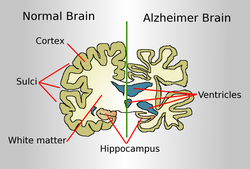
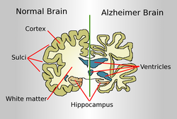
Alzheimer’s disease is a progressive, chronic disease that attacks the brain cells and neurons; dementia is the first clinical symptom of this disease, which normally afflicts people in the old age. Alzheimer’s is a serious illness that impairs the cognitive skills of the patient to such an extent that they need to be under the supervision of assisted care. Such an ailment could be understood better if scientists identify the metals of the brain, which are involved in instigating the neurodegenerative condition. Such a research study was recently completed by an international group of researchers through a collaborative approach. The research team was headed by Dr. Joanna Collingwood, who works at the School of Engineering, University of Warwick.

This research team reported that iron species in the brain could be characterized easily as they play a pivotal role in the formation of plaques caused by amyloid proteins; these proteins are extensively found in the human brain. Clustering of these proteins may reach an abnormal level, leading to the formation of plaques. The toxicity of these plaques is severe, causing apoptosis (programmed cell death) of tissues. With the progressive of tissues in the brain, the person’s cognitive skills decline consistently; memory loss is followed by deterioration in mental condition and the patient ultimately develops Alzheimer’s disease.

In patients with Alzheimer’s disease, the plaques formed by amyloid proteins are unique and can be characterized easily. The iron species are otherwise in their normal state in a healthy human brain. When a person develops Alzheimer’s disease, these iron species undergo extensive chemical reduction. This leads to the formation of a chemically reduced species named magnetite; the proliferation of magnetite occurs in the amyloid protein plaques. The team of researchers believes that magnetite is the result of the chemical interaction between iron species and amyloid proteins in the human brain afflicted with Alzheimer’s illness.

In advanced countries, like the USA and UK, there have been several advancements in diagnostic technologies used for the analysis of human brain. One such advancement is the introduction of Diamond Light Source 108 beamline instrument in Oxfordshire, UK. This sophisticated instrument is capable of performing advanced measurements of the human brain using synchrotron X-rays. The team of researchers at the University of Warwick used this instrument to gather evidences for their work. They were successful in proving that detailed chemical reduction of metallic species only took place in the human brain of individuals afflicted with Alzheimer’s disease. They further reported about the different forms of calcium species that were present in the form of minerals in the plaques formed by amyloid protein.

What exactly is the significance of this discovery? Well, we have now understood that metals in the human brain undergo a lot of chemical reduction and transformation when a person is afflicted with Alzheimer’s disease. This would help us in further discovering the root cause of the disease. By correlating the concentration of these reduced metal species with the progression of the disease, scientists can develop more innovative therapies that can exactly eradicate the root cause of the disease.

Dr. Joanna Collingwood, who is the supervisor leading this team of international researchers, works as an Associate Professor at the School Engineering, University of Warwick. She is an expert in following analytical techniques of measurement: trace metal analysis and high resolution imaging. She has used these skills to understand and elucidate the pathophysiology of neurodegenerative diseases, including Alzheimer’s disease.

According to Dr. Collingwood, we need to know the guiding principle of this research study: Iron is an essential element in the human brain, so it is but natural that we comprehensively understand how the fluctuations in iron levels could be associated with the development of Alzheimer’s disease. It is important to note that we are only concerned about iron levels in the human brain at this stage of research. We used sophisticated X-ray techniques in this study to understand how iron undergoes a step-wise change in its neurochemistry while interacting with amyloid proteins, which cluster together excessively to form plaques. It is interesting to note how amyloid protein formation instigates a massive chemical change in the composition of iron species, which are found extensively in the human brain. Previous studies have attempted to treat Alzheimer’s disease with iron-based drugs. Our findings would only alleviate further research studies in this direction, leading to the development of novel drugs based on iron.

The research study was conducted by an international collaboration between researchers working at the following institutions: University of Warwick and Keele University. It also included researchers working at the following institutions: University of Texas in San Antonio and University of Florida. To form an insight into this path-breaking discovery, the team of researchers first successfully extracted the cores formed by amyloid protein plaques; these were obtained from the human brain of two deceased patients who had succumbed to Alzheimer’s disease.

For the purpose of scanning the cores formed by amyloid plaques, researchers used the following sophisticated analytical instrument: an advanced X-ray microscope was obtained from the Advanced Light Source in Berkeley, USA. The Diamond Light Source synchrotron (beamline 108) was also used in this study; this instrument was obtained from Oxfordshire and its main purpose was to determine the chemical properties of the altered minerals found in the brain of these deceased patients. With the help of these sophisticated instruments, the researchers also analyzed whether all the iron species had undergone a change in its magnetic state when they interacted with the amyloid proteins in the plaques. Researchers tried to find out if there were any unchanged iron species in the plaques. Magnetite, the leading magnetic form of iron oxide, was found extensively in these plaques. Other minerals of altered iron species were also found in these plaques.

Significance: The UK is a small country with the best healthcare system in the world. Sadly, it still cannot cure about 850,000 patients diagnosed with dementia, the first clinical symptom of Alzheimer’s disease. The number of patients with Alzheimer’s disease is expected to be skyrocketing at 1 million and 2 million by 2025 and 2050, respectively. Presently, medical science has no cure for this disease and other conditions that cause dementia. If the development of dementia in Alzheimer’s patients is delayed by at least five years, the number of deaths caused by this condition could be halved easily. This implies that approximately 30,000 patients could be prevented from untimely death; their lives can be saved with this path-breaking discovery.

Conclusion: Researchers believe that the development and progression of Alzheimer’s disease is associated with the chemical reduction of iron species in the human brain. The resultant iron species have an altered magnetic state; the most prominent among them being magnetite. All these altered iron species are highly toxic in nature, creating conducive conditions for Alzheimer’s disease.


Bioengineering proteins for personalized medicine


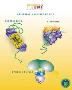
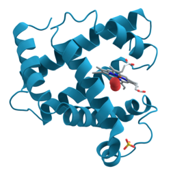
One of the latest developments in biotechnology is genetic engineering of cellular biology without the use of actual cell. This technique is termed as cell-free protein synthesis (CFPS). Chemicals, biomaterials, and medicines can be synthesized easily with this sustainable method. Cell-free systems have one major shortcoming: they cannot manufacture glycosylated proteins, that is, proteins attached to carbohydrates. There are several biological processes involving the process of glycosylation. For the prevention and treatment of diseases, it is important to understand the reaction mechanism of glycosylation. Our main purpose is to control this process and synthesize glycosylated proteins through cell-free systems.

A team of researchers have collaborated to devise a novel approach and overcome this shortcoming. The team of researchers includes following people: Dr. Matthew DeLisa, Professor of Chemical and Biomolecular Engineering at Cornell University and Dr. Michael Jewett, associate professor of chemical and biological engineering at NorthWestern University. They have devised a novel system by capitalizing on the recent advancements of CFPS technology. They have been successful in developing the missing glycosylated component through a simple reaction, which is carried out in “one-pot” system. After glycosylating the desired protein, it can be freeze-dried for later use. To use the protein for further synthesis, it can be reactivated by adding only water. The frozen protein would get thawed and retain back its natural properties at room temperature.

This team of researchers successfully published their paper titled “Single-pot Glycoprotein Biosynthesis Using a Cell-Free Transcription-Translation System Enriched with Glycosylation Machinery.” This paper was published in the latest July issue of Nature magazine. DeLisa and Jewett are the two senior lead authors of this study. According to DeLisa, they have been successful in devising the world’s first glycosylated protein through cell-free technology. This protein could be very useful in various therapeutic areas, including the development of vaccines. This is because the protein can be freeze-dried and used in various locations, indicating the portability of these protein molecules. This is a path-breaking, powerful invention that can unshackle the existing models of manufacturing proteins.

With this technology, protein-based medicine can be easily developed and transported to remote areas. Thus, the lives of several people would be saved like never before. The cost of life-saving drugs and vaccines would decrease with this novel method of synthesis. Local small-batch production of life-saving drugs can now be carried out in remote locations with low resources. Life-saving drugs have been costly till date; however, this technology aims to bring down the cost of these life-saving drugs. Therefore, poorer patients in remote areas can now have access to better healthcare.

DeLisa is a senior scientist who has spearheaded several research studies in biomedical eningeering. He has always focused on investigating the molecular mechanisms associated with the biogenesis of underlying proteins in a living cell. It is important to note that the living cell is a complex environment wherein the main barrier is the cell wall. His lab has done extensive research on several living cells, such as Escherichia coli (E.coli). According to DeLisa, it is difficult to make important breakthroughs in cell synthesis as cell walls act as barriers in the transportation of materials, including proteins. The cell wall screens all the molecules before permitting them into the cell.

Jewett works at a sophisticated biomedical laboratory in NorthWestern University. A lot research studies have been conducted into advancing the technique of cell-free synthesis, that is, efforts were made to replicate the natural biomachinery outside the cell. A collaboration between DeLisa and Jewett was nothing but fruitful in addressing their common goal: synthesis of glycosylated proteins through cell-free systems. According to Jewett, there is always a tug of war in engineering the cells of bacteria. The cell only wants to grow and survive. As a scientist, we are trying to maneuver its capability and reaction mechanisms.

To develop this novel method of synthesis, cell extracts were prepared by the team using a high quality strain of E. coli. This strain of E. coli was specifically optimized to grow in laboratory conditions. This strain of E.Coli was termed CLM24. Key components of glycosylation were used to enrich this strain of E.Coli with high selectivity. A simple reaction scheme was used to synthesize the resultant extracts. The team has christened this synthesis process as “cell-free glycoprotein synthesis (CFGpS)”

What is the unique selling point of this method? Well, the cell-free extracts obtained by this method have the complete molecular machinery required for the synthesis and glycosylation of proteins. Therefore, a molecular biologist has to simply include all DNA instructions required for the synthesis of a glycosylated protein in the desired form. Thus, CFGpS has completely broken the shackles of the existing cell-based method. Thanks to CFGpS method, we can now synthesize complex glycoproteins within a single day.

The further advantage of CFGpS method is the fact that it is highly modular in design; therefore, several varieties of glycoproteins can be easily prepared using a variety of diverse cell extracts. In this experiment, researchers used a lab-grown strain of E.coli for preparing cell extracts. It is important to note that E.coli is a simple cell, which cannot carry out glycosylation on it is own. Nevertheless, we were able to develop CFGpS platform by using this simple strain of E.coli. This implies that a completely blank slate of E.coli cells could be engineered biologically to develop into a glycosylated system of desired capacity. With this method, the structure of carbohydrates can not only be controlled but also be manipulated to suit our needs. We can synthesize highly complex glycoproteins. This was not possible till date with the existing cell-based systems.

The field of personalized medicine is growing by leaps and bounds in developed countries, including the USA. This is a very attracted protocol for on-demand drug synthesis. A simple test tube could now be used instead of a large bioreactor for drug synthesis. The whole concept of personalized medicine has received a paradigm shift with this novel method. Based on the physiology of a patient, we can now develop a unique protein molecule for drug delivery.


Can Alzheimer’s be treated with aspirin?

Plaques developed in the brain can be eliminated with a low-dose aspirin, which is an effective drug that suppresses the progression of Alzheimer’s disease. The drug aspirin is very effective in protecting the memory of patients. These are the latest findings reported by neurologists at the Rush University Medical Center. The results of this study were published in the Journal of Neuroscience.

Our study is path-breaking and novel in the sense that aspirin is one of the most commonly used medication for various illnesses. More than 1 out of 10 Americans was diagnosed with Alzheimer’s disease, which is a progressive form of dementia. Very few drugs have been approved by the FDA for the treatment of Alzheimer’s-related complications, such as dementia. Presently, only temporary relief is provided by these medications.

Researchers still do not know the exact cause of Alzheimer’s disease; however, researchers know the cause of dementia and memory loss, which is associated with the faulty disposal of amyloid beta. Amyloid beta is the most toxic protein to have been developed in the human brain. Researchers believe that the most important strategy for eliminating the progression of Alzheimer’s illness would be the activation of cellular machinery. Waste can be removed from the human brain with this machinery.

Amyloid plaques are clumps formed by the toxic protein amyloid beta. The connection between nerve cells would be harmed by amyloid plaques. Such a development is one of the major signs of Alzheimer’s illness. There seems to be a link between the reduced risk of developing Alzheimer’s disease and the consumption of aspirin. The most important component of animal cells, the lysosomes, is very useful in clearing cellular debris. In mice, lysosomes could be stimulated with aspirin. Aspirin is the component that decreases amyloid plaque.

The incidence, progression, and development of Alzheimer’s disease could be stopped by elucidating the development of amyloid plaques. To regulate the removal of waste products from the human body, a protein named TFEB. Aspirin was administered orally to mice, which were genetically modified to develop the pathology of Alzheimer’s disease.

To determine the parts of brain most affected by Alzheimer’s disease, we determined the amount of amyloid plaque in these subjects. In mice, the functions of aspirin medications are as follows: i) to augment the expression of TFEB, ii) stimulate the expression of lysosomes, and iii) decrease the pathology of amyloid plaque.

Aspirin is the most widely used medication for pain relief; moreover, it is also used extensively for the treatment of cardiovascular diseases. The findings of these research studies must be validated further. Aspirin could be soon considered as a therapeutic drug for the treatment of Alzheimer’s illness and other diseases related to dementia.


The delivery of advanced medicines is facilitated by a new technology in biomedical sciences

In Sweden, researchers at Karolinska Institutet have devised a novel technique that ensured the effective delivery of therapeutic proteins and RNA into cells. The method, which has been presented in Nature Communications, has been used to deliver gene editors and therapeutics of proteins.

The method is based on two components: the so-called extracellular vesicles and tiny bubbles, which are secreted naturally in cells. They transport molecules that are biologically active in cells. Two key components have been introduced to improve the quality of these bubbles.

These two key components are as follows: a bacterial protein called intein and a so-called fusogenic protein associated with a virus. The fusogenic protein would help the bubbles combine with the endosomal membrane and then release the contents into the cell. The intein would cut itself and would help release therapeutic proteins present inside the cell.

According to Professor Samir El Andaloussi, who is the last author of the study and researcher at the Department of Laboratory Medicine, Karolinska Institute, the engineering strategy is innovative and it presents an important step ahead in the development of extracellular vesicle technology. It can effectively overcome key barriers, which includes poor endosomal escape and a limited intracellular release.

A diverse range of conditions, such as systemic inflammation, genetic diseases, and neurological disorders have been treated with a method that has the potential of engineered EVs, which is a versatile platform that delivers therapeutics.

Dr. Xiuming Liang is the first author of this study. He has devised this technology that can effectively increase the feasibility of including advanced medicines, which improve the efficiency and reliability of therapeutic delivery in target cells.

Experiments were conducted on cells and live animals. In these studies, Cre recombinase was efficiently delivered in such a way that a protein could be cut and pasted into DNA and the Cas9/sgRNA.

These components were then used to edit genes. Cre recombinase was used to load extracellular vesicles. Then, these vesicles were injected into the brains of mice. This caused a significant change in the cells of hippocampus and cortex brain structures.

In conclusion, there is hope to use the CRISPR/Cas9 gene scissors or similar tools. These tools can treat severe genetic diseases, which occur in the central nervous system. The diseases were Huntington’s disease and spinal muscular atrophy. Systemic inflammation in mice could be treated with this technique.


The anti-tumor cells can be activated with common salt


In earlier days, cancer was supposed to be a death sentence. Presently, there have been many advancements in the treatment of cancer. Therefore, the rate of survival of cancer patients has increased, with many leading a good quality of life even after being diagnosed with cancer.

In recent years, adoptive T-cell therapy has particularly been developed as an effective cancer treatment tool. In this method, some of the body’s white blood cells, that is, the T cells, undergo such a modification that they gain the capability of recognizing and fighting tumor cells.

The efficacy of adoptive T-cell therapy depends on the metabolic activity of T cells. The T cells expression is generally suppressed in an immunosuppressive environment of tumor cells. Therefore, researchers had to identify factors that could overcome this suppression.

Historically, table salt was perceived as precious commodity. Its chemical name is sodium chloride. Today, common salt is a cheap commodity and used in kitchen recipes. A team of scientists led by Christina Zielinski have found that sodium ions, which are an integral component of common salt (sodium chloride), increased the efficiency of anti-tumor activity of T cells.

The researchers found that breast cancer tumors had a higher concentration of sodium as compared to healthy tissue. In particular, the T cells were acting strongly against tumors when the immediate environment had a high concentration of sodium.

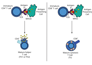
The survival time increased in such cases. Furthermore, the researchers proved that the immune response of CD8+ T cells would be enhanced by sodium.

CD8+ T cells are immune cells that identify and kill tumor cells or cells that are infected with virus in the human body. In a previous study, researchers have reported that sodium can regulate other types of T cells, which are believed to be involved in autoimmune disease and allergies.

In this study, the researchers went on to know the effect sodium could specifically have on controlling the activity of CD8+ T cells in humans.

Various technologies were used by researchers to investigate how sodium regulated the genes and the metabolic process of CD8+ T cells. The human T cells were pre-treated with salt and then they were cultured with tumors.

A mouse model of the experiment involving T cells was also carried out. The researchers reported that in the presence of salt, the metabolic fitness of CD8+ T cells improved as the uptake of sugar and amino acids increased. Thus, energy production also increased in the cells.

Consequently, the immune cells were more capable of eliminating tumor cells, as seen in the experiments of cell cultures and mouse models. The researchers found that pancreatic tumors shrank in size in mice after they were injected with T cells that were pre-treated with salt.