Researchers identify the switch that activates programmed death of cancer cells.

The UC Davis Comprehensive Cancer Center has conducted an in-depth research study. The focus was to activate programmed death of cancer cells. A crucial epitope has been identified on the CD95 receptor, and it triggers the death of cells. An epitope is a section of protein that activates the larger protein. As cell death can now be programmed, cancer treatment methods have become effective.

In molecular biology, CD95 receptors are known as Fas and death receptors. These receptors are proteins present in cell membrane. They evoke self-destruction of cells by releasing a signal. This happens when CD95 receptors are activated. By modulating the activity of Fas, researchers extended its benefits to CAR T-cell therapy. This was effective in destroying solid tumours of ovarian cancer.

Managing cancer with better therapies

The conventional method of treating cancer includes chemotherapy, radiotherapy, and surgery. In cases where cancer is diagnosed at an initial stage, these methods are effective. However, cancer cases may relapse, especially when they are therapy-resistant. Recently, CAR T-cell immunotherapy and an immune checkpoint receptor molecule have shown to activate antibodies, and thus they are promising candidates that destroy the cycle of cancerous growth.

These immunotherapeutic agents are effective against only few types of cancer cells, such as ovarian cancer, breast cancer, lung cancer, and pancreatic cancer. In CAR T-cell therapy, researchers engineer the specific type of immune cells, that is, T cells. They graft these cells on a specific antibody that targets specific tumours. The grafted T cells are quite effective in battling leukaemia and other types of blood cancer.

The engineered T cells have not been effective in combating solid tumours; the microenvironment of these tumours drives off T cells and other immune cells. Thus, they cannot provide a therapeutic effect to solid tumours. Although the immune receptor activates antibodies, the T cells cannot infiltrate without additional spaces.

The activity of death receptors

Now, let us understand the activity of death receptors. Through targeted therapy, we can trigger them into programming cell death of tumours. Thus, chemotherapeutic drugs should be such that they induce the activity of death receptors. Many pharmaceutical companies have been slightly successful in targeting the death receptor-5. But the clinical trials of Fas agonists have failed.

Developing the right target

The activity of immune cells is effectively regulated by Fas. However, researchers have proposed that cancer cells can be targeted selectively if they identify the correct epitope. After identifying the targeted epitope, researchers of this study have designed a new type of antibodies. These antibodies show selectivity while binding and activating Fas. With this strategy, specific tumor cells can be destroyed.


Researchers develop a remote-controlled cancer immunotherapy system


An innovative ultrasound system has been developed to destroy genetically controlled processes in live T cells of the immune system. This team of researchers can destroy cancer cells. By developing non-invasive immunotherapeutic strategies, cancer cells can be manipulated and destroyed.

A novel strategy was used to improve the practical applications of mechanogenetics, which is a scientific discipline that improves the expression of genetics and activity of cells. T cells were mechanically destroyed by ultrasound. To genetically control cells, mechanical signals were used.

This experimental study establishes how mechanogenetics system is remote controlled and T cells are manipulated by chimeric antigen receptor (CAR). Cancer cells can be targeted and killed with this innovative approach. Researchers have modified CAR-T cells with mechano-sensors, genetically transducing modules.

This innovative approach was termed as therapy of CAR-T cells, which provided a paradigm shift for the treatment of cancer. Life-threatening complications develop when CAR-T cells are non-specifically targeted. The precision and the accuracy of CAR-T cell specific immunotherapy was improved in an unprecedented manner.

This innovative immunotherapy was used to target solid tumors. At the same time, off-tumor activities were minimized. Microbubbles were combined with streptavidin and they were attached to cell surface. Mechanical vibration and the stimulation of Piezo1 ion specific channels was performed by microbubbles when they were exposed to waves of ultrasound.

This led to the entry of calcium ions into the cell, triggering the following downstream pathways: the activation of calcineurin, the dephoshorylation of NFAT and the translocation into the nucleus. With recognition and destruction of targeted cancer cells, chimeric antigen receptor (CAR) was used to initiate the expression of genes.


Cancer stem cells can now be destroyed by targeting metabolism

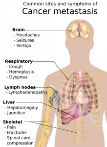
Cancer is a fatal illness with poor prognosis and survival rate, especially when it has progressed to a metastatic state. Scientists have not yet been able to decipher why patients become resistant to chemotherapeutic drugs and therapies. To address this objective, researchers worked diligently at  the Rogel Cancer Center—it is affiliated to the University of Michigan.

They made an important breakthrough in the year 2003. The lead supervisor was Dr S. Wicha, MD for the team of researchers. They found that there are cancer stem cells that act like a fuel within a tumor. Although this group of cells is immensely small, they are the ones that trigger the growth and metastasis of cancer.

The simple strategy was then to simply kill the group of cancer stem cells, and the long lost battle against cancer could be defeated easily. But, is this so easy to sound hopeful for cancer patients? Not really, cancer is such a condition that can relapse and attack patients even after they have been cured temporarily.

Currently, there has been an important discovery: cancer stem cells do not really exist in ONLY a single state but they are exhibited in different states; they are immensely plastic in nature. This implies that different forms can be easily adopted by cancer stem cells.

They could be in a dormant state for some point of time and then easily bounce back into uncontrolled growth, leading to formation of tumor. Multiplication and spreading, the two characteristic features of cancer stem cells, have been attributed to its most important property: plasticity.

Presently, patients are treated with targeted therapies for combating cancer. Although these therapies are effective, they have been successful in destroying tumor cells only for a certain period of time. There are many cases in which patients develop resistance to these targeted therapies.

What is the cause of drug resistance in cancer patients? Most scientists believe that drug resistance is triggered once again by cancer stem cells. Because cancer stem cells have high plasticity, they change their form completed when subjected to targeted therapies.

The resultant effect is that cancer stem cells are completely unrecognizable to these therapies following change of form. The patient thus develops resistance to therapies and the patients’ condition deteriorates consistently.

The conclusion: multiple stem cell therapies must be developed to effectively combat every form of cancer stem cell. This is a humungous task to achieve according to scientists at the Rogel Cancer Center. Cell metabolism is the key feature that controls the plasticity of cancer stem cells.

How do we eliminate the plasticity of cancer stem cells? Well, all we need to do is to target the metabolism of cancer stem cells. In other words, cancer stem cells can be effectively attacked by destroying cell metabolism.Mitochondria are cell organelles that supply energy to cells, irrespective of its kind. This includes cancer stem cells.

Mitochondria are organelles that perform cellular respiration, depending completely on the supply of oxygen. Cells derive energy from mitochondria, which converts sugar or glucose molecules into energy with the help of cellular oxygen.

Cancer stem cells are very unique due to its plasticity. When they are in the dormant state, they derive energy from glucose molecule. When they grow in a proliferative state, cancer stem cells depend completely on oxygen. Given the mechanism of deriving energy for sustenance and proliferation, researchers attacked both forms of cell metabolism observed in cancer stem cells.

They used a drug that is conventionally used for treating arthritis. This drug can effectively block the functioning of mitochondria in cancer stem cells. The levels of cellular glucose were further manipulated to obstruct the pathway of energy. They performed this experiment on cancer-stricken mice.

To their surprise, they had effectively knocked off all the cancer stem cells from the mice. This is an important breakthrough in cancer research, and the findings of this study have attracted a lot of attention. The complete experiment has been published in Cell Metabolism, a peer-reviewed SCI journal.

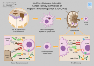
The general public may wonder why this study is so path-breaking and innovative in nature. Well, the conventional cancer therapy makes use of highly toxic chemicals to destroy cells in a tumor. Here, researchers adopted a completely different pathway to control the explosion of cancer stem cells: they destroyed the cell metabolism associated with the proliferation of tumor cells.

According to the lead researcher Dr. Wicha, further studies must be conducted to understand how metabolism controls the efficacy of human immune system. This could open a new chapter in cancer research: scientists could then focus their efforts on developing novel combinatorial techniques for cancer treatment.

These techniques must aim at effectively combining existing immunotherapies with anti-stem cell therapies. The concept is refreshing and offering new hope; however, extensive clinical trials must be conducted to validate results.


New methods approved by FDA for treating digestive tract cancers

The drug Lutathera (lutetium Lu 177 dotatate) was approved by US FDA for treating  neuroendocrine tumors that originate in the gastrointestinal tract and pancreas (GEP-NETs). This is an important breakthrough as it is the first radioactive drug to have been approved by the FDA. It is now a novel treatment for GEP-NETs.

Lutathera drug was found to be quite effective in treating adult patients with GEP-NETs. The treatment options for GEP-NETs were limited as it is a rare type of cancer and the conventional therapy was not successful in preventing the proliferation of this cancer. With US FDA approving the drug Lutathera, it is a ray of hope for patients diagnosed with these rare type of cancer.

It also establishes that US FDA is now open to considering data from alternative therapies, which can provide hope to patients with this rare type of cancer. GEP-NETs develop not only in the pancreas but also in different parts of the digestive system, such as stomach, colon, intestine, and rectum.

Statistical data suggests that the diagnosis of GEP-NETs has been low each year. Only one out of 27,000 people develop this cancer in the United States of America. Lutathera is a radiopharmaceutical drug. It exerts its therapeutic activity by clinging to the somatostatin receptor, which is a component of cell and is present in certain types of tumors.

After clinging to  the receptor, the drug gets into the cell and exudes radiation to damage cancerous activity. The drug Lutathera was approved by two research studies: a randomized clinical trial was conducted on 229 patients, who were diagnosed with a certain type of advanced GEP-NET. These patients elicited a positive response to the drug.

In this clinical trial, a combination of Lutathera and octreotide drug was administered to some patients, while the remaining patients only received   octreotide drug. After providing treatment to patients, researchers measured the period of time for which tumors did not show any signs of grow or development. This period was defined as progression-free survival of patients.

Patients who consumed a combination of Lutathera and octreotide drugs had a longer period of progression-free survival as compared to patients who only consumed the drug octreotide. Moreover, the possibility of the growth of tumors or death was lower in patients who received both the drugs: Lutathera and octreotide .

The second research study was performed on 1,214 patients in Netherlands. These patients were diagnosed with tumors showing positive response to somatostatins, including GEP-NETS. The drug Lutathera was administered at a single site in these patients.

In a clinical trial that included 360 patients with GEP-NETs, it was found that tumors  either shrunk completely or partially in about 16 percent of included patients . These patients also received the drug Lutathera initially, and their responses were monitored by the US FDA.

The drug Lutathera has following side-effects: the white blood cells decrease sharply in patients (lymphopenia); the levels of enzymes become high in certain organs (increased GGT, AST and/or ALT); some patients may develop nausea and vomiting; there could be a sudden spike in blood sugar levels (hyperglycemia), and the levels of potassium become low in the blood (hypokalemia).

The drug Lutathera has following serious side-effects: blood cells may decrease sharply in numbers (myelosuppression); certain types of blood or bone marrow cancers may develop in patients(secondary myelodysplastic syndrome and leukemia);  some patients may witness a serious damage to their kidneys (renal toxicity); some patients may suffer from permanent liver damage (hepatotoxicity); the hormonal levels may become abnormal in the human body (neuroendocrine hormonal crises), and infertility.

The drug Lutathera can harm the fetus developing in the womb of a pregnant mother; therefore, pregnant women are educated about the potential damage caused to the fetus by this drug . In general, radiation exposure is provided to patients before administering the drug.