Verapamil: an effective therapy for type 1 diabetes

To promote the functioning of beta cells and insulin in patients with type 1 diabetes, researchers have developed a novel strategy that minimizes the requirements of insulin and the incidence of hypoglycemia. These researchers have worked at the Comprehensive Diabetes Center at the University of Alabama in Birmingham, USA.

The journal Nature Medicine published these findings recently. Verapamil is the most commonly used drug for controlling blood pressure; it was approved for oral administration in the year 1981. Following the administration of verapamil, type 1 diabetes patients were able to produce greater amounts of insulin. Thus, their daily requirement of insulin was reduced substantially and their blood sugar levels were also in control.

The drug verapamil is not just safe and effective for type 1 diabetes but also a promising therapy that provides new hope to people living with this life-threatening condition. These were positive results confirmed in a human clinical trial that was randomized and double-blinded in nature; the clinical trial was controlled by placebo.

Verapamil has shown promising results in improving the function of beta cells in pancreas; the functioning of beta cells is related to the control of insulin production. Optimum levels of insulin ensure a good quality of life in patients. Under such a scenario, type 1 diabetes patients have new hope. It is otherwise difficult to control such a life-threatening condition that offers no hope.

Although this drug does not sound like a complete cure for type 1 diabetes, it is however a promising therapy for altering a life-threatening condition: type 1 diabetes. Such patients need to boost the production of insulin in their body in order to have better disease control.

A clinical trial was conducted on animal models in the year 2014. In this clinical trial, it was reported that the condition of type 1 diabetes could be completely reversed by administering verapamil. Then, they conducted a human clinical trial to determine the effects of this drug.

For more than three decades, the drug verapamil has been approved by the FDA for the treatment of high blood pressure. Current research findings are path-breaking in the sense that the drug is quite safe and effective in the treatment of type 1 diabetes patients. In patients with type 1 diabetes, the body’s immune system attacks beta cells of the pancreas.

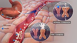
These beta cells are responsible for the production of insulin. Insulin is the hormone that controls blood sugar levels in the patient. The production of insulin decreases substantially when the beta cells of pancreas are destroyed in the human body.

Consequently, blood sugar levels would rise in the human body and the patient would become extremely dependent on external sources for insulin. The function of beta cells can be preserved effectively when a patient is administered verapamil.

This drug induces the body to produce more insulin. In various clinical trials, it has been proved that the participants’ dependency on external insulin decreases substantially. Several individuals with type 1 diabetes can effectively regulate their blood sugar levels with this strategy.

In the human clinical trial, the drug verapamil was administered to 24 patients. These patients were in the age group of 18 to 45 years. Over the course of one year, verapamil was administered to 11 patients while a placebo drug was administered to 13 patients.

Only patients with type 1 diabetes were included in this clinical trial. They received insulin therapy to manage their condition throughout the duration of this clinical trial. The total daily dose of insulin was monitored in both the groups, that is, the group that received verapamil and the group that received placebo.

Moreover, we also monitored the amount of insulin produced in these groups. Factors such as the percentage of change in insulin and HbA1C levels were also monitored. There were patients who experienced hypoglycemic events; all such events of each patient were recorded in our human clinical trial.

A continuous glucose monitoring system was used to determine the healthy blood glucose levels of each patient. Patients with type 1 diabetes do have therapeutic options for hope. In fact, such patients should be able to deal with the illness in a promising way following the successful administration of verapamil.

Insulin dependency was substantially reduced in patients with type 1 diabetes following the administration of verapamil. The quality of life was significantly improved in these patients. The risk of comorbidities would be improved when the overall blood sugar levels was controlled in patients. Thus, a patient with type 1 diabetes would not develop several other comorbidities, such as kidney disease, blindness, and heart attack.


Researchers develop a remote-controlled cancer immunotherapy system


An innovative ultrasound system has been developed to destroy genetically controlled processes in live T cells of the immune system. This team of researchers can destroy cancer cells. By developing non-invasive immunotherapeutic strategies, cancer cells can be manipulated and destroyed.

A novel strategy was used to improve the practical applications of mechanogenetics, which is a scientific discipline that improves the expression of genetics and activity of cells. T cells were mechanically destroyed by ultrasound. To genetically control cells, mechanical signals were used.

This experimental study establishes how mechanogenetics system is remote controlled and T cells are manipulated by chimeric antigen receptor (CAR). Cancer cells can be targeted and killed with this innovative approach. Researchers have modified CAR-T cells with mechano-sensors, genetically transducing modules.

This innovative approach was termed as therapy of CAR-T cells, which provided a paradigm shift for the treatment of cancer. Life-threatening complications develop when CAR-T cells are non-specifically targeted. The precision and the accuracy of CAR-T cell specific immunotherapy was improved in an unprecedented manner.

This innovative immunotherapy was used to target solid tumors. At the same time, off-tumor activities were minimized. Microbubbles were combined with streptavidin and they were attached to cell surface. Mechanical vibration and the stimulation of Piezo1 ion specific channels was performed by microbubbles when they were exposed to waves of ultrasound.

This led to the entry of calcium ions into the cell, triggering the following downstream pathways: the activation of calcineurin, the dephoshorylation of NFAT and the translocation into the nucleus. With recognition and destruction of targeted cancer cells, chimeric antigen receptor (CAR) was used to initiate the expression of genes.


Cancer stem cells can now be destroyed by targeting metabolism

Cancer is a fatal illness with poor prognosis and survival rate, especially when it has progressed to a metastatic state. Scientists have not yet been able to decipher why patients become resistant to chemotherapeutic drugs and therapies. To address this objective, researchers worked diligently at  the Rogel Cancer Center—it is affiliated to the University of Michigan.

They made an important breakthrough in the year 2003. The lead supervisor was Dr S. Wicha, MD for the team of researchers. They found that there are cancer stem cells that act like a fuel within a tumor. Although this group of cells is immensely small, they are the ones that trigger the growth and metastasis of cancer.

The simple strategy was then to simply kill the group of cancer stem cells, and the long lost battle against cancer could be defeated easily. But, is this so easy to sound hopeful for cancer patients? Not really, cancer is such a condition that can relapse and attack patients even after they have been cured temporarily.

Currently, there has been an important discovery: cancer stem cells do not really exist in ONLY a single state but they are exhibited in different states; they are immensely plastic in nature. This implies that different forms can be easily adopted by cancer stem cells.

They could be in a dormant state for some point of time and then easily bounce back into uncontrolled growth, leading to formation of tumor. Multiplication and spreading, the two characteristic features of cancer stem cells, have been attributed to its most important property: plasticity.

Presently, patients are treated with targeted therapies for combating cancer. Although these therapies are effective, they have been successful in destroying tumor cells only for a certain period of time. There are many cases in which patients develop resistance to these targeted therapies.

What is the cause of drug resistance in cancer patients? Most scientists believe that drug resistance is triggered once again by cancer stem cells. Because cancer stem cells have high plasticity, they change their form completed when subjected to targeted therapies.

The resultant effect is that cancer stem cells are completely unrecognizable to these therapies following change of form. The patient thus develops resistance to therapies and the patients’ condition deteriorates consistently.

The conclusion: multiple stem cell therapies must be developed to effectively combat every form of cancer stem cell. This is a humungous task to achieve according to scientists at the Rogel Cancer Center. Cell metabolism is the key feature that controls the plasticity of cancer stem cells.

How do we eliminate the plasticity of cancer stem cells? Well, all we need to do is to target the metabolism of cancer stem cells. In other words, cancer stem cells can be effectively attacked by destroying cell metabolism.Mitochondria are cell organelles that supply energy to cells, irrespective of its kind. This includes cancer stem cells.

Mitochondria are organelles that perform cellular respiration, depending completely on the supply of oxygen. Cells derive energy from mitochondria, which converts sugar or glucose molecules into energy with the help of cellular oxygen.

Cancer stem cells are very unique due to its plasticity. When they are in the dormant state, they derive energy from glucose molecule. When they grow in a proliferative state, cancer stem cells depend completely on oxygen. Given the mechanism of deriving energy for sustenance and proliferation, researchers attacked both forms of cell metabolism observed in cancer stem cells.

They used a drug that is conventionally used for treating arthritis. This drug can effectively block the functioning of mitochondria in cancer stem cells. The levels of cellular glucose were further manipulated to obstruct the pathway of energy. They performed this experiment on cancer-stricken mice.

To their surprise, they had effectively knocked off all the cancer stem cells from the mice. This is an important breakthrough in cancer research, and the findings of this study have attracted a lot of attention. The complete experiment has been published in Cell Metabolism, a peer-reviewed SCI journal.

The general public may wonder why this study is so path-breaking and innovative in nature. Well, the conventional cancer therapy makes use of highly toxic chemicals to destroy cells in a tumor. Here, researchers adopted a completely different pathway to control the explosion of cancer stem cells: they destroyed the cell metabolism associated with the proliferation of tumor cells.

According to the lead researcher Dr. Wicha, further studies must be conducted to understand how metabolism controls the efficacy of human immune system. This could open a new chapter in cancer research: scientists could then focus their efforts on developing novel combinatorial techniques for cancer treatment.

These techniques must aim at effectively combining existing immunotherapies with anti-stem cell therapies. The concept is refreshing and offering new hope; however, extensive clinical trials must be conducted to validate results.


Bioengineering proteins for personalized medicine

One of the latest developments in biotechnology is genetic engineering of cellular biology without the use of actual cell. This technique is termed as cell-free protein synthesis (CFPS). Chemicals, biomaterials, and medicines can be synthesized easily with this sustainable method. Cell-free systems have one major shortcoming: they cannot manufacture glycosylated proteins, that is, proteins attached to carbohydrates.

There are several biological processes involving the process of glycosylation. For the prevention and treatment of diseases, it is important to understand the reaction mechanism of glycosylation. Our main purpose is to control this process and synthesize glycosylated proteins through cell-free systems.

A team of researchers have collaborated to devise a novel approach and overcome this shortcoming. The team of researchers includes following people: Dr. Matthew DeLisa, Professor of Chemical and Biomolecular Engineering at Cornell University and Dr. Michael Jewett, associate professor of chemical and biological engineering at NorthWestern University.

They have devised a novel system by capitalizing on the recent advancements of CFPS technology. They have been successful in developing the missing glycosylated component through a simple reaction, which is carried out in “one-pot” system. After glycosylating the desired protein, it can be freeze-dried for later use. To use the protein for further synthesis, it can be reactivated by adding only water. The frozen protein would get thawed and retain back its natural properties at room temperature.

This team of researchers successfully published their paper titled “Single-pot Glycoprotein Biosynthesis Using a Cell-Free Transcription-Translation System Enriched with Glycosylation Machinery.” This paper was published in the latest July issue of Nature magazine. DeLisa and Jewett are the two senior lead authors of this study.

According to DeLisa, they have been successful in devising the world’s first glycosylated protein through cell-free technology. This protein could be very useful in various therapeutic areas, including the development of vaccines. This is because the protein can be freeze-dried and used in various locations, indicating the portability of these protein molecules. This is a path-breaking, powerful invention that can unshackle the existing models of manufacturing proteins.

With this technology, protein-based medicine can be easily developed and transported to remote areas. Thus, the lives of several people would be saved like never before. The cost of life-saving drugs and vaccines would decrease with this novel method of synthesis.

Local small-batch production of life-saving drugs can now be carried out in remote locations with low resources. Life-saving drugs have been costly till date; however, this technology aims to bring down the cost of these life-saving drugs. Therefore, poorer patients in remote areas can now have access to better healthcare.

DeLisa is a senior scientist who has spearheaded several research studies in biomedical eningeering. He has always focused on investigating the molecular mechanisms associated with the biogenesis of underlying proteins in a living cell.

It is important to note that the living cell is a complex environment wherein the main barrier is the cell wall. His lab has done extensive research on several living cells, such as Escherichia coli (E.coli). According to DeLisa, it is difficult to make important breakthroughs in cell synthesis as cell walls act as barriers in the transportation of materials, including proteins. The cell wall screens all the molecules before permitting them into the cell.

Jewett works at a sophisticated biomedical laboratory in NorthWestern University. A lot research studies have been conducted into advancing the technique of cell-free synthesis, that is, efforts were made to replicate the natural biomachinery outside the cell.

A collaboration between DeLisa and Jewett was nothing but fruitful in addressing their common goal: synthesis of glycosylated proteins through cell-free systems. According to Jewett, there is always a tug of war in engineering the cells of bacteria. The cell only wants to grow and survive. As a scientist, we are trying to maneuver its capability and reaction mechanisms.

To develop this novel method of synthesis, cell extracts were prepared by the team using a high quality strain of E. coli. This strain of E. coli was specifically optimized to grow in laboratory conditions.

This strain of E.Coli was termed CLM24. Key components of glycosylation were used to enrich this strain of E.Coli with high selectivity. A simple reaction scheme was used to synthesize the resultant extracts. The team has christened this synthesis process as “cell-free glycoprotein synthesis (CFGpS)”

What is the unique selling point of this method? Well, the cell-free extracts obtained by this method have the complete molecular machinery required for the synthesis and glycosylation of proteins.

Therefore, a molecular biologist has to simply include all DNA instructions required for the synthesis of a glycosylated protein in the desired form. Thus, CFGpS has completely broken the shackles of the existing cell-based method. Thanks to CFGpS method, we can now synthesize complex glycoproteins within a single day.

The further advantage of CFGpS method is the fact that it is highly modular in design; therefore, several varieties of glycoproteins can be easily prepared using a variety of diverse cell extracts. In this experiment, researchers used a lab-grown strain of E.coli for preparing cell extracts. It is important to note that E.coli is a simple cell, which cannot carry out glycosylation on it is own.

Nevertheless, we were able to develop CFGpS platform by using this simple strain of E.coli. This implies that a completely blank slate of E.coli cells could be engineered biologically to develop into a glycosylated system of desired capacity. With this method, the structure of carbohydrates can not only be controlled but also be manipulated to suit our needs.

We can synthesize highly complex glycoproteins. This was not possible till date with the existing cell-based systems. The field of personalized medicine is growing by leaps and bounds in developed countries, including the USA. This is a very attracted protocol for on-demand drug synthesis.

A simple test tube could now be used instead of a large bioreactor for drug synthesis. The whole concept of personalized medicine has received a paradigm shift with this novel method. Based on the physiology of a patient, we can now develop a unique protein molecule for drug delivery.


Can Alzheimer’s be treated with aspirin?

Plaques developed in the brain can be eliminated with a low-dose aspirin, which is an effective drug that suppresses the progression of Alzheimer’s disease. The drug aspirin is very effective in protecting the memory of patients. These are the latest findings reported by neurologists at the Rush University Medical Center. The results of this study were published in the Journal of Neuroscience.

Our study is path-breaking and novel in the sense that aspirin is one of the most commonly used medication for various illnesses. More than 1 out of 10 Americans was diagnosed with Alzheimer’s disease, which is a progressive form of dementia. Very few drugs have been approved by the FDA for the treatment of Alzheimer’s-related complications, such as dementia. Presently, only temporary relief is provided by these medications.

Researchers still do not know the exact cause of Alzheimer’s disease; however, researchers know the cause of dementia and memory loss, which is associated with the faulty disposal of amyloid beta. Amyloid beta is the most toxic protein to have been developed in the human brain. Researchers believe that the most important strategy for eliminating the progression of Alzheimer’s illness would be the activation of cellular machinery. Waste can be removed from the human brain with this machinery.

Amyloid plaques are clumps formed by the toxic protein amyloid beta. The connection between nerve cells would be harmed by amyloid plaques. Such a development is one of the major signs of Alzheimer’s illness. There seems to be a link between the reduced risk of developing Alzheimer’s disease and the consumption of aspirin. The most important component of animal cells, the lysosomes, is very useful in clearing cellular debris. In mice, lysosomes could be stimulated with aspirin. Aspirin is the component that decreases amyloid plaque.

The incidence, progression, and development of Alzheimer’s disease could be stopped by elucidating the development of amyloid plaques. To regulate the removal of waste products from the human body, a protein named TFEB. Aspirin was administered orally to mice, which were genetically modified to develop the pathology of Alzheimer’s disease.

To determine the parts of brain most affected by Alzheimer’s disease, we determined the amount of amyloid plaque in these subjects. In mice, the functions of aspirin medications are as follows: i) to augment the expression of TFEB, ii) stimulate the expression of lysosomes, and iii) decrease the pathology of amyloid plaque.

Aspirin is the most widely used medication for pain relief; moreover, it is also used extensively for the treatment of cardiovascular diseases. The findings of these research studies must be validated further. Aspirin could be soon considered as a therapeutic drug for the treatment of Alzheimer’s illness and other diseases related to dementia.


New treatment strategy for kidney cancer

Kidney cancer is a potentially lethal illness with little hope for cure. At the University of North Carolina, scientists have been trying to explore a new treatment therapy for kidney cancer. The Lineberger Comprehensive Cancer Center is attached to the University of North Carolina.

Scientists working at this institute have developed a potential therapeutic target that can be used for treating kidney cancers; they have been successful in identifying the gene that causes kidney cancer.

An overabundance of blood vessels leads to tremendous genetic change in patients with kidney cancer. Owing to the excessive flow of blood, tumors are developed easily. This finding is promising enough to be considered as a pathway for the development of cancer in patients.

A genetic change was observed in more than 90 percent of patients, which were diagnosed with the most common type of kidney cancer.  It is important to note that VHL is a tumor suppressor gene, which is lost due to a change in genetic conditions.

In these cells, there is an over-accumulation of a protein termed as ZHX2. The over-accumulated protein would instigate other signals, which are involved in the growth of cancerous tumors. Based on these findings, we suggest that ZHX2 is potentially a new therapeutic target that is associated with the development of renal cell carcinoma.

Following the suppression of the gene VHL, several ZHX2 proteins would be accumulated in the human body. Consequently, signals related to kidney cancer would be promoted. The expression of this protein must be destroyed in order to treat kidney cancer patients; the therapeutic treatment may be a single drug or combination of drugs.

Genetic mutations or alterations have occurred in more than 90 percent of cases with renal cell carcinoma.In patients with renal cell carcinoma, VHL is the most important gene that suppresses tumor.

Several reports have suggested that VHL plays an important role in every stage of renal cell carcinoma, which includes initiation to tumor progression to metastasis.

It is important to note how kidney cancer would be developed with the loss of function of VHL. In kidney cancer patients, the downstream effects of VHL function loss can be targeted therapeutically.

Several cell signals are involved in the excessive production of blood vessels. There are FDA approved drugs that block these signals, which cause downstream manifestation of VHL protein. This would be the standard mode of treatment for patients with renal cell carcinoma.

Most patients would hardly respond to these drugs. Moreover, these patients would quite often show drug resistance; therefore, researchers wanted to identify other targets that were accumulated in cells that lacked normal functioning of VHL gene. Cancerous cell growth was promoted with the abnormal functioning of VHL gene.

Researchers wanted to understand how oncogenesis is being promoted in kidney cancer cells following the loss of VHL function. A screening technique was created by researchers to identify new molecules, which would be useful in driving cancer cells after the loss of function of VHL.

In kidney cancer cells, the expression of VHL was lacking but the expression of ZHX2 was promoted. From laboratory models, the protein ZHX2 was eliminated completely. With this treatment strategy, the growth of cancer cells would be inhibited. Moreover, metastasis of cancer would also be suppressed effectively.

Several novel therapies have been developed for the treatment of kidney cancer. These therapies are as follows: i) molecular target therapy and ii) treatments based on immunology. However, several novel therapeutic targets must be used to treat metastatic condition in patients with kidney cancer.

Mutated forms of VHL are observed in most patients with kidney cancer; therefore, it is very imperative to investigate this gene. There have been several advancements in kidney cancer treatment modalities in the past few years. More than a dozen drugs have been approved by the FDA for the treatment of kidney cancer.


In severe cases of COVID-19, autoantibodies cause havoc and even death


It is a well-known fact that the pandemic of COVID-19 can be controlled only by boosting the production of antibodies in the immune system of patients. However, a recent article in Nature magazine reports otherwise. According to scientists at Yale University, the immune system of patients with severe COVID-19 is impaired significantly and boosting the production of antibodies cannot really control the illness.

Diseases like lupus and rheumatoid arthritis are autoimmune disorders, which are treated by boosting the production of autoantibodies. These antibodies interact and target damaged tissues associated with autoimmune diseases. In patients afflicted with COVID-19, autoantibodies target multiple organ systems that are otherwise healthy. This means that healthy tissues in the brain, liver, and gastrointestinal tract are attacked by autoantibodies. Moreover, they also target blood vessels and platelets in the bloodstream. This implies that the severity of COVID-19 is directly proportional to the number of detected autoantibodies.

Scientists at Yale University also found out that these autoantibodies attack many proteins in the immune system, that is, proteins that otherwise effectively fight infections are damaged by autoantibodies. Therefore, the situation is like a double-edged sword in patients with severe COVID-19 infection. In general, antibodies are effective in combating infection, but when COVID-19 infection is severe, patients develop autoantibodies. These autoantibodies attack multiple types of cells and tissues.

In most cases of COVID-19, the infection became severe and led to the production of autoantibodies, which are antibodies that caused extensive damage. However, it must be noted that in most severe cases of COVID-19, patients already had a pre-existing disease that caused the production of autoantibodies. The observation was done after testing mice with pre-existing autoimmune disorders and who had developed COVID-19 infection. Such mice had a large concentration of autoantibodies. Such kind of sickly mice were more likely to succumb to COVID-19 infection as there is no effective cure till date.

Autoantibodies are also called rogue antibodies and are believed to be existing for a long period of time in patients with pre-existing autoimmune disorders. When such patients contracted COVID-19 infection, they developed severe form of the disease that could not be tackled with existing medications and injections. The medical symptoms of COVID-19 became severe and long lasting in these patients. In other words, the virus became a legacy in the bodies of these patients. Therefore, all patients with autoimmune disorder should immediately be vaccinated to prevent more cases of severe COVID-19.

In patients with autoimmune disorder, autoantibodies are produced even at a mild stage of COVID-19 infection. The study was conducted by an esteemed team of scientists and physicians working at Yale University. In their clinical practice, they made concerted efforts to tackle COVID-19 infection: they screened blood samples of 194 patients with COVID-19 infection; the extent of severity was different in different patients. Nevertheless, autoantibodies were detected in all the blood samples of these patients.

Yale scientists developed a novel technology to detect the extent of damage caused by autoantibodies to proteins of the immune system. The technology was named Rapid Extracellular Antigen Profiling (REAP), and it explored the interaction of autoantibodies with approximately 3,000 proteins of the human body.

The scientists at Yale believe that the study’s findings may be used to develop novel strategies to combat or even prevent the damage caused by autoantibodies in patients with severe infection of COVID-19. Moreover, REAP technology is not just restricted to measuring the response of autoantibodies to COVID-19 infection.

The technology can be used to determine the damage caused by autoantibodies in patients with many types of autoimmune disorders and chronic diseases. These scientists are now exploring whether the technology can be used to determine the damage caused by autoantibodies in cancer patients and in patients with neurological disorders.


How to standardize metrics and indicators of the quality of research papers


Thanks to advances in computer algorithms and data science, several metrics and indicators are today available for evaluating the quality of research papers. The impact of a research paper can be evaluated with these metrics that are specially designed to suit the needs of an individual researcher, a team of researchers, a department of an institution, etc.

Peer review is a qualitative process, but metrics is a quantitative parameter and the choice of metrics depends on the goal and subject of a research study. Despite receiving funding for metrics research, scientists still do not have high quality metrics to evaluate the findings of a research study that presents global data. Despite having shortcomings, journal level indicators are still being used as parameters for research quality evaluation.

A 2014 conference on Science and Technology Indicators (STI) was held at Leiden in Netherlands. The panel consisted of eminent professors and professionals from a leading STM publisher, Elsevier in Netherlands. They felt that there is an urgent need to standardize the quality of metrics and indicators that are used to evaluate the content and data presented in research studies.

For this purpose, they coined the term “Snowball Metrics” initiative: all international universities decided to collaborate and develop a set of standards for evaluating the quality of research in terms of both output and research methodology. This is a collaborative effort between the Director of Research Metrics at Elsevier and eminent professors of leading universities in the world. The moot points of Elsevier’s vision are as follows:

  • An entire workflow of a research study is evaluated on various aspects with the help of several metrics.
  • Peers related to the subject of a research study must be allowed to select their set of metrics.
  • It should be possible to generate and use metrics in an automated fashion and at a scalable level.
  • In general, the focus of a research study is to answer a question related to science, technology, or medicine. A metrics evaluates the data of the study in quantitative fashion, but this data should also be validated qualitatively with evidences to ensure that the findings of the research study are complete.
  • The quantitative inputs of multiple metrics are found to be most reliable in nature.
  • Metrics are affected by some characteristics, such as discipline and so they must be considered even if they do not reflect the performance of a research study.
  • The use of metrics can be done in a transparent way only when researchers do not use manipulative ways of “gaming.” Nevertheless, researchers still use metrics in an irresponsible and incorrect way.
  • Researchers who use metrics on a daily basis should be given the responsibility of defining the set of metrics that they need everyday for use. Thus, this community of researchers can alone come forth with the definitions of various metrics.
  • There should never be any black boxes in research methodologies.
  • An aggregation of metrics should never be considered. Moreover, composite metrics never reflect the true value of data presented in a research study.
  • The methodologies used to evaluate the quality of metrics should never be related to the sources of data and the tools used to generate the data. Moreover, the business models and access codes used to gather underlying data should never be affected by metrics.


Pfizer signs a cooperation deal with Gilead for remdesivir, a COVID 19 medication


Pfizer signed a cooperation deal with Gilead Sciences to produce and supply remdesivir, a promising drug used to treat COVID-19. According to this agreement, Pfizer shall provide production services at its McPherson, Kansas factory in the US. The company would be manufacturing and supplying the drug remdesivir for Gilead.

After signing the cooperation agreement, Pfizer became a promising company in the fight against COVID-19. The deal is aligned with the “innovation ecosystem” that commits to fight a battle against COVID-19 pandemic. The agreement has brought about a fruitful collaboration between small biotech companies and large pharmaceutical companies. Many government agencies associated with academia would be benefitted from this cooperation.

The COVID-19 pandemic has become a real menace, and a single company cannot really bring about an innovation and put an end to the COVID-19 menace. The deal between Pfizer and Gilead would be good enough to bring about an innovative ecosystem, which will deliver effective medical solutions. Both companies would be working together to manufacture vaccines, sterile injections, and biologics.


300 million doses of COVID-19 vaccine to be supplied to European Union by Sanofi and GSK

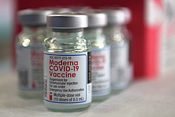
The European Union would be receiving upto 300 million doses of COVID-19 vaccine by GSK and Sanofi. Sanofi and GSK collaborated with each other to produce the vaccine candidate, which is based on recombinant protein-based technology and adjuvant technology. European countries like France, Belgium, Germany, and Italy would be manufacturing doses of this vaccine. This is a major breakthrough in fighting against COVID-19.

This is the first major vaccine against COVID-19 and it will be benefit one million Europeans, provided it is safe and effective at the clinical trial stage. The vaccine would be beneficial in tackling the global pandemic of coronavirus.

In a press release statement, the President of GSK vaccines thanked Sanofi for collaborating with them, and reiterated that the vaccine would be available soon in Europe. Both companies have worked hard to scale up operations and introduce a vaccine that will be able to tackle the growing pandemic of COVID-19.

The clinical trial would be conducted by Sanofi, and the phase 3 stage of the trial would be completed by 2020. If the vaccine produces positive results, the team of scientists at Sanofi would be filing for a regulatory approval, which is expected to be completed by the first quarter of 2021. Moreover, Sanofi and GSK have collaborated together to produce an antigen and adjuvant that would produce about one billion doses per year.

Globally, the vaccine would be available through a collaborative effort by Sanofi and GSK

In the United States and the UK, Sanofi and GSK have signed cooperation agreement with the Biomedical Advanced Research and Development Authority. The companies have decided to scale up the production of the vaccine at a global level by the end of 2021/22. The collaboration would include governments and global health organizations.